Molecular Analysis of Dinosaur Osteocytes Indicates Presence of Endogenous Molecules
October 22, 2012

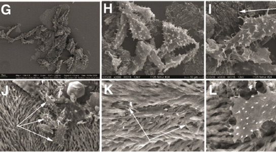
G: Under SEM, a mass of cells from MOR 1125 is seen to be closely associated with demineralized matrix. H: Higher magnification shows that all cells have remnants of filipodia, and demonstrate wide variation in shape. I: Single osteocyte from T.rex showing elongate morphology and longer filipodia in some areas of the cell (arrows). J: Three clustered osteocytes (arrows) on fibrous matrix of MOR 2958. K: Single osteocyte within lacunae of fibrous matrix. Filipodia can be seen entwining with fibrous matrix (arrows). L: Large osteocyte and matrix. Image: Schweitzer et al.
20 years ago, Mary Schweitzer discovered that she spotted the effects of what could only be described as a red blood cell in a slice of dinosaur bone. This seemed impossible, since organic remains weren’t supposed to be able to survive the fossilization process. Numerous tests indicated that the spherical structures were blood cells from a 67-million year old Tyrannosaurus rex.
In the following years, Schweitzer and her colleagues discovered more soft tissues, which could be either blood vessels or feather fibers. However, skeptics have been arguing that the organic tissues were simply microbes that had invaded the fossilized bone.

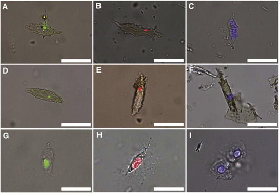
A: T.rex; D, B. canadensis; G: Ostrich cells showing small localized region of binding of anti-DNA antibodies interior to the cell membrane. Reactivity of antibodies to ostrich cells is enhanced, consistent with the presence of a greater quantity of immunoreactive material in these extant cells. B: Trex; E: B. canadensis; and H, ostrich osteocytes showing positive response to propidium iodide (PI), a DNA intercalating dye, to a similar small region of material internal to cell. C: T.rex; F: B. canadensis; and I, ostrich cellular response to 4′,6′-diamidino-2-phenylindole dihydrochloride (DAPI), another DNA-specific stain.
Schweitzer and her colleagues have continued to amass support and the latest evidence comes from a molecular analysis of osteocytes from T. rex andBrachylophosaurus canadensis. The cells were isolated and when exposed to an antibody that targets a protein, the cells reacted as expected. And when the dinosaur cells were subjected to more tests involving other antibodies that target DNA, the antibodies bound to material in small, specific regions inside the apparent cell membrane.
Mass spectrometry uncovered amino acid sequences of proteins in the extracts of dinosaur bones that matched the sequences from actin, tubulin and histone4 that are present in the cells of all animals. Some microbes have similar proteins, but tests showed that soil-derived E. coli as well as sediments surrounding the two dinosaur specimens failed to bind actin and tubulin antibodies, which bound to the extract containing the osteocytes. Schweitzer and her collaborators published their findings in the journal Bone and also presented them in Raleigh, at the annual meeting of the Society of Vertebrate Paleontology.
No comments:
Post a Comment